Amyloid β-protein “Amyloid fibril” extends under the microgravity environment – Flash analysis report of the first “Amyloid” space experiment handled by Astronaut Kanai –
Last Updated:
July 19, 2018
National Research and Development Agency
Japan Aerospace Exploration Agency (JAXA)
Exploratory Research Center on Life and Living Systems (ExCELLS)/
Institute for Molecular Science (IMS),
Inter-University Research Institute Corporation
National Institutes of Natural Sciences (NINS)
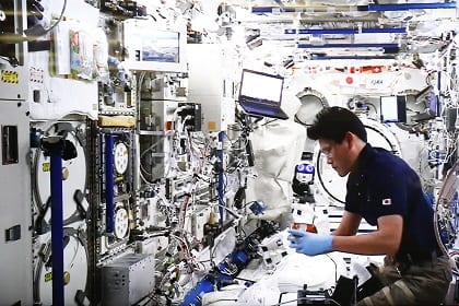
In January 2018, the first Amyloid Experiment to examine the formation mechanism of amyloid fibril (amyloid β: Aβ), considered as a factor of the onset of Alzheimer’s disease, by using the microgravity environment of Japanese Experiment Module “Kibo” of the International Space Station (ISS) . Astronaut Kanai was in charge of the experiment operation.
The first Amyloid Experiment was intended to prepare amyloid fibril with small amount of amyloid β-protein solution in different conditions by using the incubator of the Cell Biology Experiment Facility (CBEF) . Amyloid fibrils formed from amyloid β protein crystals were retrieved by four separate times, respectively, six hours, a day, three days, and nine days after the experiment started, and kept frozen. The frozen experiment samples were safely returned to Earth aboard the US Dragon spacecraft and are being under analysis at the National Institutes of Natural Sciences (NINS).
First, the formation of amyloid fibrils was examined.
Fluorescent dyes were added to experiment samples before the experiment started. The fluorescent dyes, which does not develop colors before amyloid β-protein forms amyloid fibrils, bind to amyloid fibrils and develop colors only when amyloid fibrils were formed. This property enables the extension rate of amyloid fibrils to be grasped by examining the intensity of color development of the fluorescent dyes.
The frozen experiment samples which were retrieved separately, six hours, a day, three days, and nine days after the experiment were defrosted on the ground after their return and their fluorescence intensity was measured.
Consequently, the measurement found that amyloid fibrils were formed from amyloid β-protein under the microgravity environment.
According to the results of the preceding experiments showing that protein crystals grow more slowly under the microgravity environment, it was assumed that amyloid fibril might not be formed. However, the experiment revealed that amyloid fibrils are formed from amyloid β-protein under the microgravity environment, just as shown by the hypothesis.
Subsequently, the extension rate of amyloid fibrils was examined.
For comparison, amyloid fibril formation was also attempted on the ground with experiment samples stored on the ground, the same as those used in space. The experiment samples in orbit equipped with a small thermometer in advance were monitored for their temperature changes in orbit. By reproducing the same temperature conditions as in orbit based on the monitoring information, 1G experimental samples were placed in the incubator, retrieved separately, six hours, a day, three days, and nine days after starting the experiment, and kept frozen. As in orbit, the fluorescent measurement was performed to compare the result in orbit and that on the ground. The comparison revealed that amyloid β-protein extends more slowly in orbit than on the ground. The more slow extension of amyloid fibril in orbit has added to the potential for the formation of high-quality amyloid fibril under the microgravity environment, which cannot be obtained on the ground.
The second Amyloid experiment is intended to prepare more amount of amyloid-β protein solution after the conditions and kinds of samples are narrowed down based on the first experiment result, to extend amyloid fibril under the microgravity environment, and to examine the structure of amyloid fibril with an electronic microscope or other tools after collecting and analyzing the collected samples.

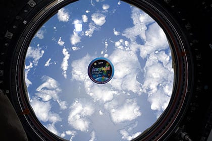
Decal for Amyloid experiment photographed against the window of “Kibo”
(January 2018)(Credit: JAXA/NASA)
*All times are Japan Standard Time (JST)

Comments are closed.